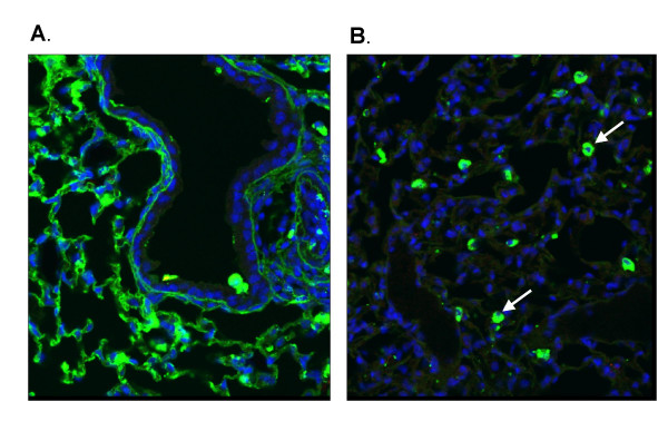
Figure 5.
Distribution of FITC-labelled siRNA and asODN in mouse lung. FITC-labelled asODN (a) and siRNA (b) (160 μg/mouse) were complexed to GL67 and "sniffed" into mouse lung. One or 24 hours after transfection the lungs were paraffin-embedded and processed for confocal microscopy. Nuclei are shown in blue, FITC signal is shown in green. Arrow indicates alveolar macrophage.

