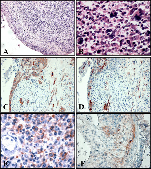
Figure 1.

• A: Cystic area delimited by benign squamous epithelium, with transitional area to premalignant. Sarcomatous component is present. • B: Pleomorphic cells in sarcomatous component. • C: Positive staining for CK AE1/AE3 in squamous epithelial lining and squamous infiltrating component. • D: Positive staining for alpha smooth muscle actin in basal atypical cells (for control look at positivity in vessels). • E: Positive staining for alpha smooth muscle actin in sarcomatous cells. • F: Positive staining for CD10 in epithelial basal cells and stromal component.
