
Figure 1.
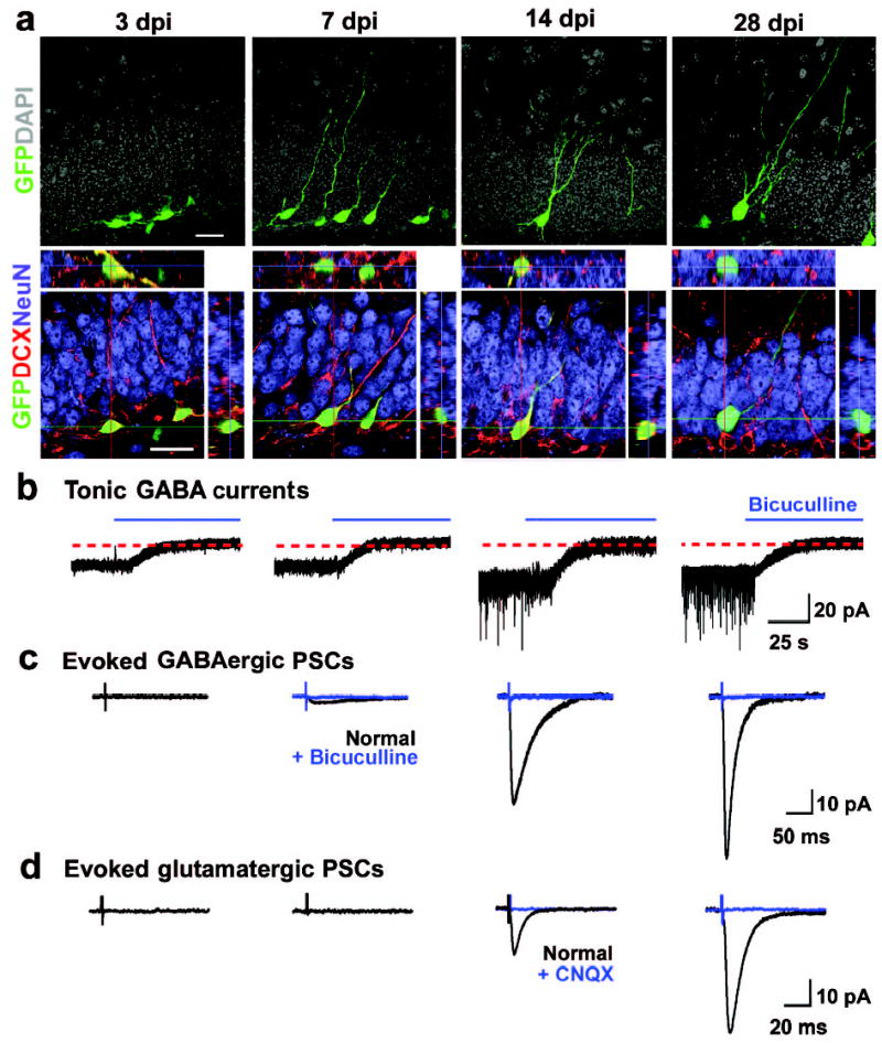
Development of newborn DGCs in the adult mice. a, Confocal images of new DGCs (GFP+, green) at different stages. Shown are projections (top) and confocal images of immunostaining (bottom) for doublecortin (DCX, red) and NeuN (blue) with orthogonal views to confirm the co-localization of GFP and DCX or NeuN. Scale bars: 20 μm. b–d, Synaptic integration of newborn DGCs. Shown are sample recording traces from GFP+ DGCs under whole-cell voltage-clamp (Vm = −65 mV). Tonic currents shown are continuous recordings before and after adding bicuculline (100 μM, blue). Evoked PSCs shown are averaged responses from 5 consecutive stimuli before (black) and after (blue) adding bicuculline (10 μM) or CNQX (50 μM), as indicated. Scale bars: 20 pA and 25 s (b); 10 pA and 50 ms (c); 10 pA and 20 ms (d).