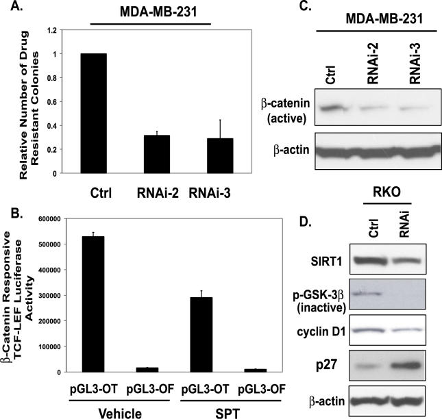
Figure 6. SIRT1 Inhibition Affects Key Phenotypic Aspects of Cancer Cells.
(A) MDA-MB-231 cells were infected for two rounds with RNAi-2 and −3 retrovirus, and puromycin-resistant colonies were counted after 3 d of selection. Error bars indicate standard deviation from the average of three experiments.
(B) RKO cells were transfected with 500 ng of pGL3-OT, a TCF-LEF−responsive reporter, or pGL3-OF, a negative control with a mutated TCF-LEF binding site in combination with 10 ng of pRL-CMV vector. Twenty-four hours post-transfection, cells were treated with either vehicle (DMSO) control or with 700 μM SPT for 24 h. Firefly luciferase activity was measured and normalized to the Renilla luciferase activities.
(C) As described in (A), pooled populations of MDA-MB-231 cells stably expressing RNAi-2 or RNAi-3 were harvested, protein concentrations were determined, and Western blot analysis was performed. An antibody that specifically recognizes the unphosphorylated (active) form of β-catenin was used, and on the same blot, β-actin was probed to ensure equal loading.
(D) Western blot analysis was performed on RKO cells expressing control or SIRT1 RNAi. Antibodies against SIRT1, phospho-GSK3β (inactive), cyclin D1, p27, and β-actin were used for Western blotting. On the same blot, β-actin was probed to ensure equal loading.

