
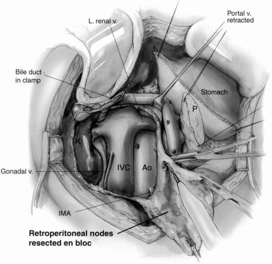
Figure 2. The retroperitoneal dissection component of the radical procedure. The retroperitoneum is dissected from the hilum of the right kidney (K) to the left lateral border of the aorta (Ao) in the horizontal axis, exposing the left renal vein. In the vertical axis, the dissection extends from the level of the portal vein to below the level of the third portion of the duodenum (level of the inferior mesenteric artery [IMA] origin). Here, the gastric staple line and pancreatic remnant (P) are being retracted toward the upper right. The inferior vena cava (IVC) and aorta are fully exposed, and the right gonadal vein has been preserved. A curved vascular clamp gently occludes the inferior aspect of the bile duct. The retroperitoneal fat and lymph nodes are being resected en bloc (bottom right).