
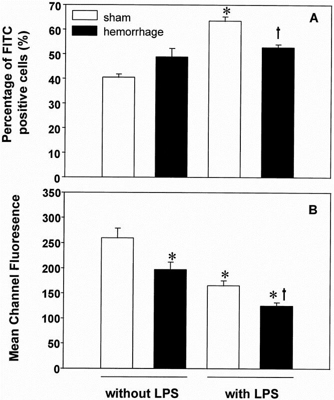
Figure 6. Percentage of FITC-positive staining wound exudate cells (A) and the mean channel fluorescence of positive-staining cells (B) harvested on the first postoperative day and stimulated with or without 10 μg/ml lipopolysaccharide A for 1 hour. FITC-positive cells represent cells that have incorporated fluorescent S. aureus BioParticles that had been incubated together with the wound exudate cells for 30 minutes. Analysis of variance, *p < 0.05 vs. sham mice without stimulation, †p < 0.05 vs. sham mice with stimulation.