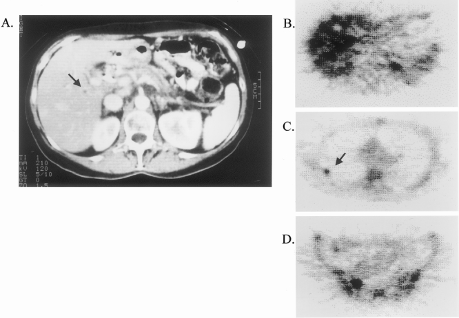
Figure 2. 18FDG-PET documentation of CT-occult metastatic disease. The patient had obstructive jaundice. CT demonstrated a potentially resectable primary lesion in the pancreatic head, with an indeterminate subcentimeter lesion in the right lobe of the liver (A, arrow). 18FDG-PET demonstrated multiple CT-occult hepatic metastases (B), as well as unsuspected skeletal metastases in rib (C, arrow) and pelvis (D).
