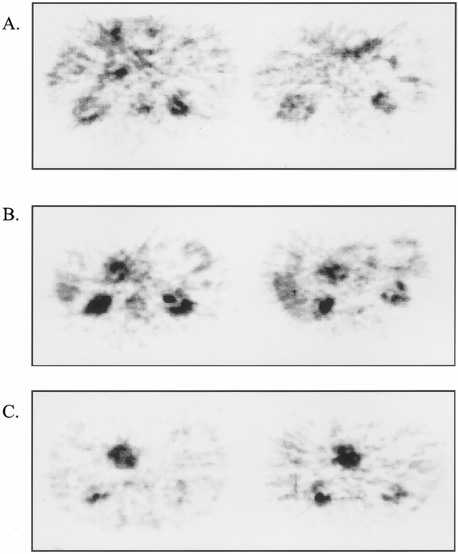
Figure 3. Assessment of response to chemoradiation using 18FDG-PET. Three representative examples of patients with biopsy-documented pancreatic ductal adenocarcinoma assessed by 18FDG-PET before (left panels) and after (right panels) neoadjuvant chemoradiation. In all three patients, CT showed no change in measurable tumor diameter. (A) Patient with treatment response documented by 18FDG-PET (pretreatment SUR = 3.0, posttreatment SUR = 0.0); note physiologic uptake of 18FDG in stomach anteriorly and in kidneys bilaterally. The resected specimen demonstrated grade 2A treatment effect (30% necrosis). (B) Patient with stable disease (<50% reduction in SUR) documented by 18FDG-PET (pretreatment SUR = 3.8, posttreatment SUR = 3.2). Resected specimen showed grade 1 treatment effect (<10% tumor necrosis). (C) Patient with progressive disease documented by 18FDG-PET (pretreatment SUR = 3.5, posttreatment SUR = 5.6). Disease was unresectable at laparotomy because of local tumor extension.
