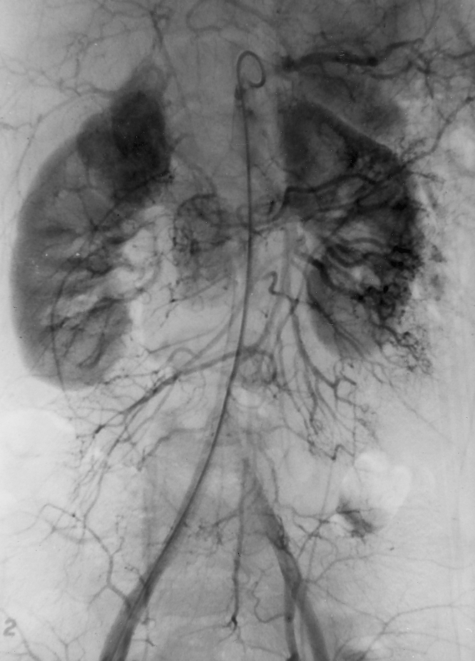
Figure 1. Angiogram demonstrating the typical radiographic blush of an extraadrenal pheochromocytoma to the patient’s right of the aorta. This is also the angiogram that did not detect a second extraadrenal pheochromocytoma located on the patient’s left side; it was successfully resected at a second operation.
