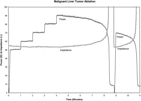
Figure 2. A graphic display of power (in watts) and tissue impedance (in ohms) during RFA of a malignant liver tumor. Using ultrasound guidance, the LeVeen needle electrode is placed into the tumor and the multiple array is deployed. Treatment is then initiated at 50 W of power and increased by 10 W at 1-minute intervals up to a maximum power of 90 W. At 8 minutes, RFA-induced coagulative necrosis of the tumor is occurring; the tissue impedance at this point rapidly rises to more than 200 ohms and the power output precipitously falls (roll-off). After waiting 20 seconds to allow heat in the tumor to dissipate, RF energy is again applied at approximately 75% (70 W) of the maximum power achieved, until tissue impedance again rises and power rolls off. This two-phase application of RF energy is performed in each area treated with the LeVeen multiple array needle electrode.

An official website of the United States government
Here's how you know
Official websites use .gov
A
.gov website belongs to an official
government organization in the United States.
Secure .gov websites use HTTPS
A lock (
) or https:// means you've safely
connected to the .gov website. Share sensitive
information only on official, secure websites.
