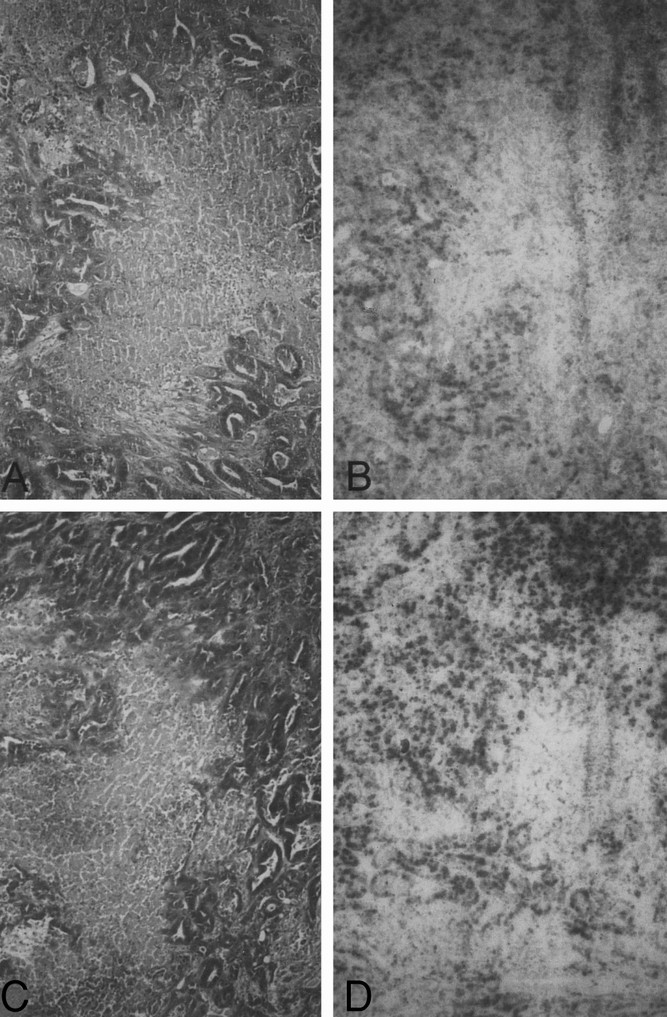
Figure 3. Histologic findings in group II (DSM alone). (A) Microscopic section of tumor tissue showing irregularly distributed, indirect signs of irreversible cell damage 24 hours after treatment. Tumor cells display clear shrinkage and partial loss of cell contact. The cell nuclei are pyknotic and basophilic and the cytoplasm is eosinophilic. There were central, large and partially confluent necrotic areas comprising completely homogenized, eosinophilic tumor cells (bar = 2 μm, H&E). (B) BrdU incorporation was focally detected in the peripheral tumor 24 hours after therapy (bar = 5 μm, BrdU histochemistry). (C) Histologic picture 14 days after treatment is similar to that 24 hours after treatment (bar = 2 μm, H&E). (D) Microscopic section 14 days after treatment shows strong BrdU incorporation of the tumor cells. The eosinophilic tumor necrosis never demonstrated BrdU incorporation (bar = 5 μm, BrdU histochemistry).
