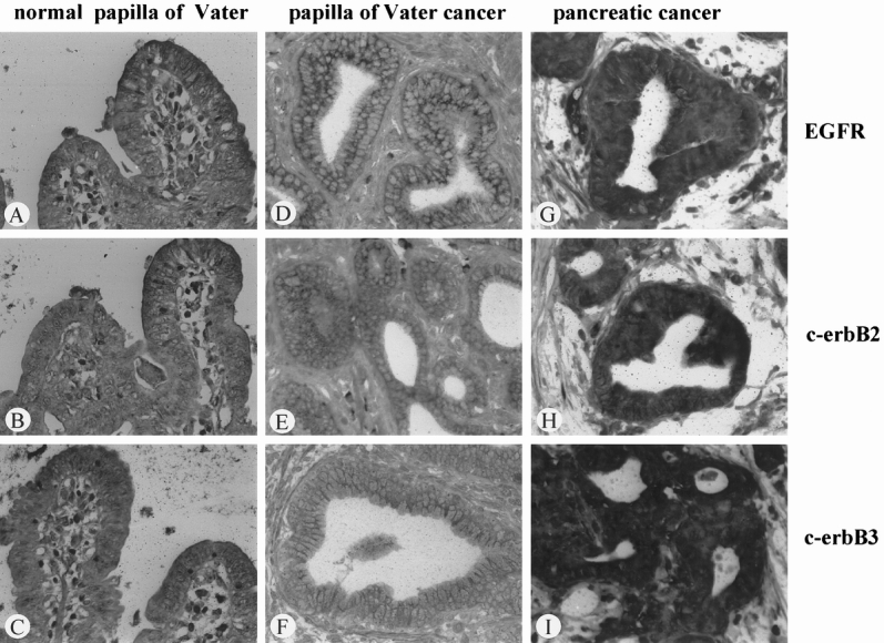
Figure 2. In situ hybridization of EGFR (A, D, G), c-erbB2 (B, E, H), and c-erbB3 mRNA (C, F, I) in normal papilla of Vater tissue samples (A through C), papilla of Vater cancer (D through F), and pancreatic cancer (G through I). Whereas in pancreatic cancer the in situ hybridization signals were markedly increased in comparison with normal tissues, in papilla of Vater cancer samples the mRNA signals were in general weaker than in normal tissues. (Original magnification ×300)
