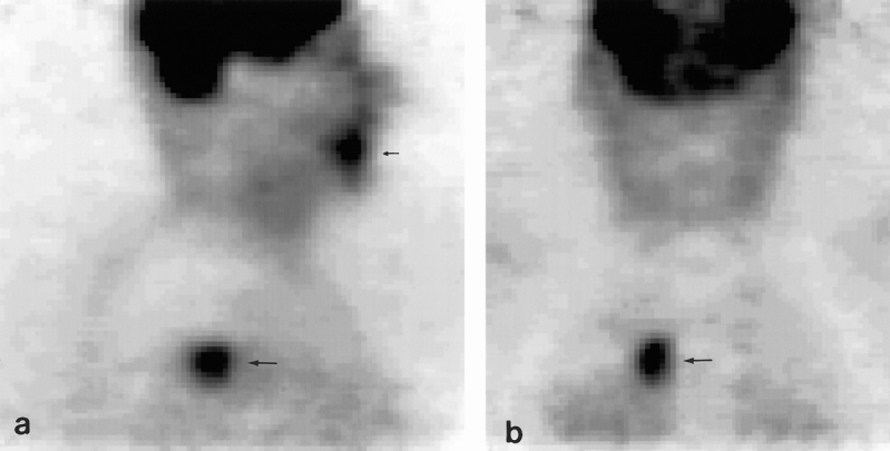
Figure 1. Sagittal (A) and coronal (B) slices of an FDG dual-head positron emission tomography study in a patient with a squamous cell carcinoma of the gingiva, demonstrating increased uptake at that site (small arrow). Unexpectedly, increased FDG accumulation was also seen in the upper lobe of the right lung (large arrow). Histologic examination of the biopsy specimen revealed an adenocarcinoma; therefore, this tumor was classified as an unknown second primary tumor.
