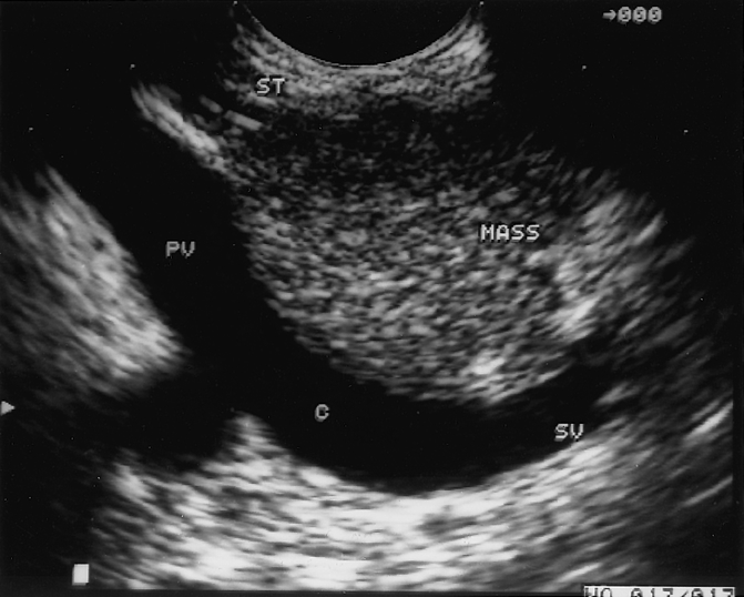
Figure 3. Mass in the head of the pancreas secondary to chronic pancreatitis. This image was obtained by endoscopic ultrasound and demonstrates a mass in the head of the pancreas adjacent to the portal vein. The patient underwent a Whipple resection, resulting in complete resolution of pain. PV, portal vein; SV, splenic vein; C, confluence of splenic and superior mesenteric veins.
