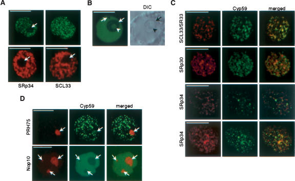
FIGURE 6.
Subnuclear localization of AtCyp59. (A) Cofocal images of nuclei from tobacco cells expressing AtCyp59-GFP (single confocal section, left; maximum intensity projection of all sections of the same nucleus, right) and two SR proteins, SRp34 and SCL33 (two lower panels; shown are only single confocal sections). Note that AtCyp59-GFP localizes into punctuate pattern, which is different from speckled pattern of SR proteins. Arrows point to nucleoli. Bars, 8 μm. (B) Single confocal section of Arabidopsis nucleus expressing AtCyp59-GFP (left) with corresponding DIC image (right). Arrows point to nucleoli and arrowhead to nucleolar cavity. Bar, 8 μm. (C) Colocalization studies of AtCyp59 with Arabidopsis SR proteins. Tobacco protoplasts were transiently cotransformed with plasmids expressing AtCyp59-GFP and indicated SR proteins fused to RFP. Maximum intensity projections of cotransformed nuclei are shown. For SRp34–AtCyp59 combination also a single confocal section is shown (second row from bottom). Merged images show superimposition of GFP and RFP signals. Bars, 7 μm. (D) Colocalization studies of AtCyp59 with markers for nucleoli [PRH75-RFP (tobacco protoplasts) and Nop10-RFP (Arabidopsis protoplasts)]. Only single confocal sections are shown. Arrows point to nucleoli. Bars, 5 μm.

