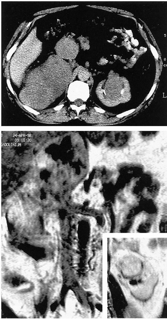
Figure 1. Patient 12. Abdominal computed tomography scan (top) and nuclear magnetic resonance imaging (bottom) showing a large right retroperitoneal mass involving the retrohepatic inferior vena cava and pushing the right kidney forward (not shown), and a synchronous mass in the sinus of the left kidney. In this difficult case, the patient was informed of the possible need for bilateral nephrectomy, but conservation of the right kidney turned out to be possible.
