
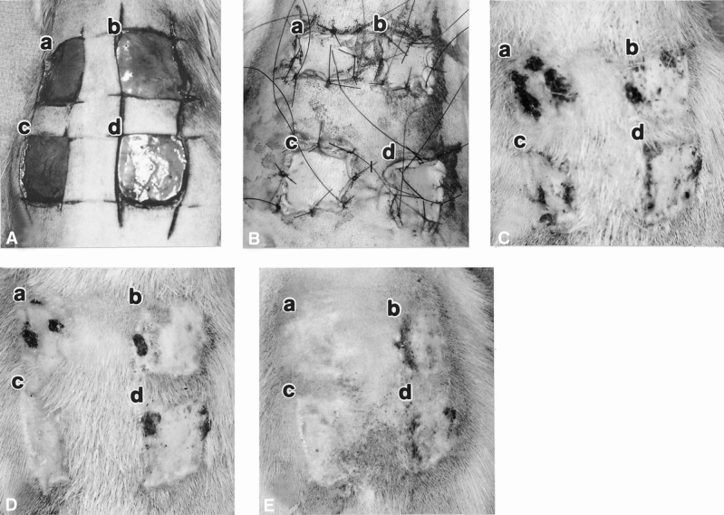
Figure 1. Macroscopic appearance of full-thickness wounds on rat dorsum implanted with acellular dermal matrix (ADM) with immediate onlay split-thickness autografts (STSGs) on day of surgery showing open full-thickness wounds (A), closed wounds containing implants (B), day 14 (C), day 20 (D), and day 30 (E). In each picture: (a) xenogenic ADM with STSG (0.005 inches thick), (b) STSG control (0.005 inches thick), (c) allogenic ADM with STSG (0.005 inches thick), (d) STSG control (0.017 inches thick). Dark areas on the wounds are regions of graft necrosis.