
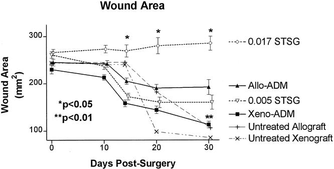
Figure 3. Areas for wounds implanted with porcine acellular dermal matrix (ADM) and split-thickness autografts (STSG; Xeno-ADM), rat ADM and STSG (Allo-ADM), thin STSG control (0.005 STSG), thick STSG control (0.017 STSG), untreated allograft (rejection control), and untreated xenograft (rejection control) groups. The wound area of the xenogenic ADM was significantly less (** P < .01, paired t test) than that of the 0.017 STSG and allogenic ADM groups on day 30. The wound area of the 0.017 STSG was significantly greater (* P < .05, paired t test) than the Allo-ADM, 0.005 STSG, and Xeno-ADM. Shown are means ± standard error (n = 29).