
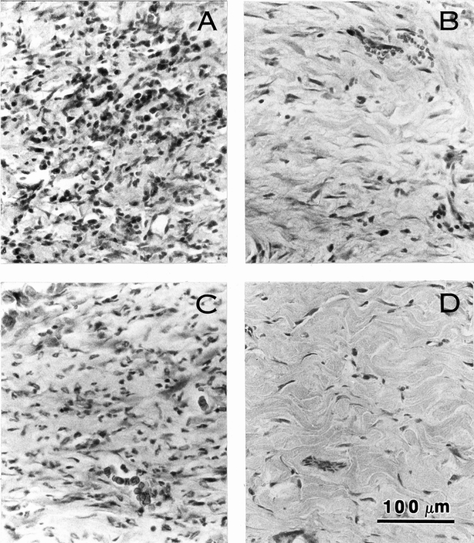
Figure 7. Microscopic appearance of acellular dermal matrix (ADM) with onlay split-thickness skin grafts (STSG) in full-thickness wounds in rat on postoperative day 20. Extensive inflammatory infiltrate is seen in the sample from the wound treated with xenogenic ADM with STSG (A) and limited inflammation is seen in samples from wounds implanted with thin STSG (B) or allogenic ADM and thin STSG (C). No inflammation or tissue disruption is seen in the thick STSG (D) group. Hematoxylin and eosin stain. Magnification bar = 100 μm.