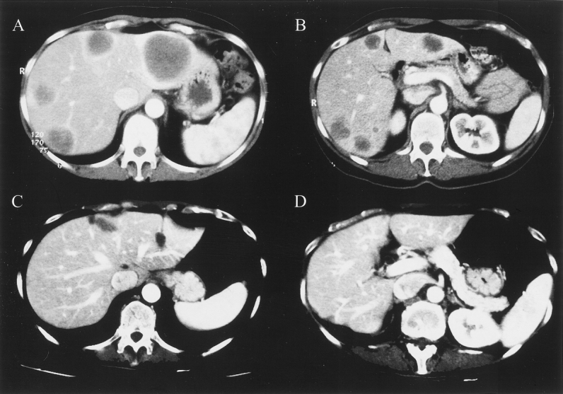
Figure 3. Comparative abdominal computed tomographs of patient 13, treated by a two-stage hepatectomy. Liver sections (A, B) illustrate the multinodular bilobar lesions before the first hepatectomy (12 metastases). (C, D) Liver after multiple partial resection. This patient was free of recurrence 7 months after the two-stage hepatectomy.
