
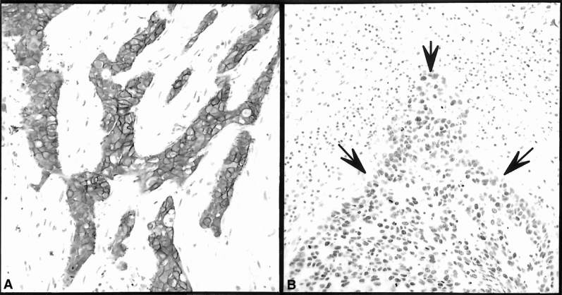
Figure 1. Immunohistochemical determination of c-erbB-2 (A) and p53 (B) expression in breast cancer and adjacent normal-appearing tissue. There is c-erbB-2 (A) membrane immunostaining of the malignant cells shown. This microscopic section was evaluated based on the intensity of staining and the percentage of malignant cells with membrane staining and was scored as 3/3. There is an increase in p53 (B) staining of the malignant cells demarcated by the solid arrows. This microscopic section was evaluated based on the intensity of staining and the percentage of malignant cells staining and was scored as 3/3 and 4/5, respectively (overall score 7).