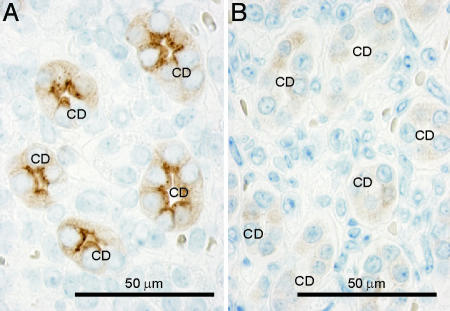
Fig. 3.
Immunohistochemical labeling of AQP2 in control (A) and AQP2-total-KO (B) mice. In kidneys from control mice, strong apical labeling is associated with CD principal cells (CD). In contrast, weak and diffuse cytoplasmic labeling was observed in kidney CD principal cells in AQP2-total-KO mice, indicating the presence of low levels of truncated AQP2ΔE3 protein.

