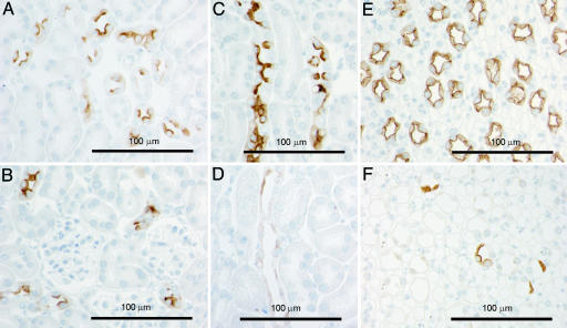
Fig. 4.
Immunohistochemical labeling of AQP2 in sections of kidneys from control (A, C, and E) and AQP2-CD-KO (B, D, and F) mice. A and B are from kidney cortex displaying CNTs. C and D are from kidney outer medulla displaying outer medullary CDs, and E and F are from kidney inner medulla displaying inner medullary CD. The CNT segment in the AQP2-CD-KO mice (B) expresses wild-type AQP2 protein.

