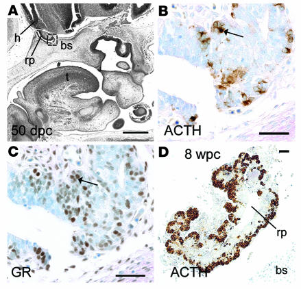
Figure 7. Human early anterior pituitary development.
(A) Sagittal section from head at 50 dpc. Pound symbol indicates oral cavity. (B–D) Brightfield IHC with antibodies to ACTH (B and D) and GR (C) counterstained by toluidine blue. B and C show higher-magnification views of boxed region in A. Arrows show overlapping expression profiles of cytoplasmic ACTH and nuclear GR in adjacent sections. (D) Anterior pituitary at 8 wpc. bs, basosphenoid bone; h, developing hypothalamus; rp, Rathke’s pouch; t, tongue. Scale bars: 500 μm (A), 100 μm (B and C), 200 μm (D).

