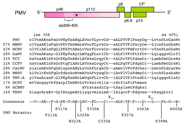
Figure 4.
Replicase motif conserved in the Tombusviridae. The PMV genome map as shown in Figure 1. The amino acids (aa) 306–405 (speckled region) represent a conserved domain (CD), common to the analogous Tombusviridae proteins, as determined by BLAST-PSI and manual alignment. The percent identities for this region are indicated on the left and the virus abbreviations are defined in the Methods. The amino acids are given in single-letter code. Alanine-scanning mutagenesis was targeted to ten amino acids on the PMV genome selected from the consensus sequence.

