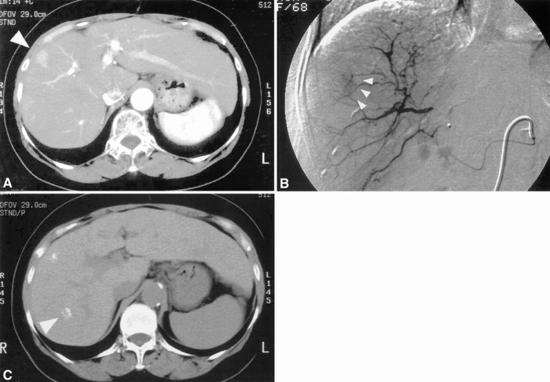
Figure 2. A 68-year-old woman in whom an hepatocellular carcinoma (HCC) nodule was correctly identified by Lipiodol computed tomography (CT) but not by helical CT. (A) Helical CT scan during the arterial phase shows a 1.5-cm hypervascular tumor in S8 (arrowhead). (B) Digital subtraction angiography confirmed the helical CT findings (arrowheads). (C) Lipiodol deposits were identified in S7 (arrowhead) in addition to S8. Both lesions were confirmed to be HCC by intraoperative ultrasound and postoperative histologic evaluation.
