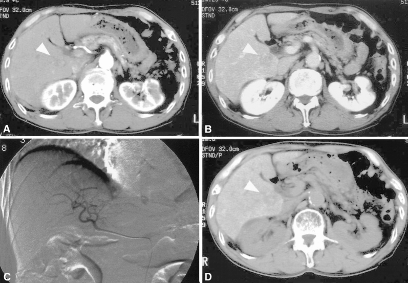
Figure 3. A 78-old-year man in whom helical computed tomography (CT) identified an hepatocellular carcinoma nodule not detected by Lipiodol CT. (A, B) A nodule measuring 2.3 cm in diameter (arrowhead) was depicted in the caudate lobe as a low-density lesion during both the arterial (A) and the portal venous (B) phase by helical CT. (C) This lesion was not detected by digital subtraction angiography. (D) No Lipiodol deposit was seen in the lesion (arrowhead). This lesion was confirmed to be well-differentiated hepatocellular carcinoma by postoperative histologic examination.
