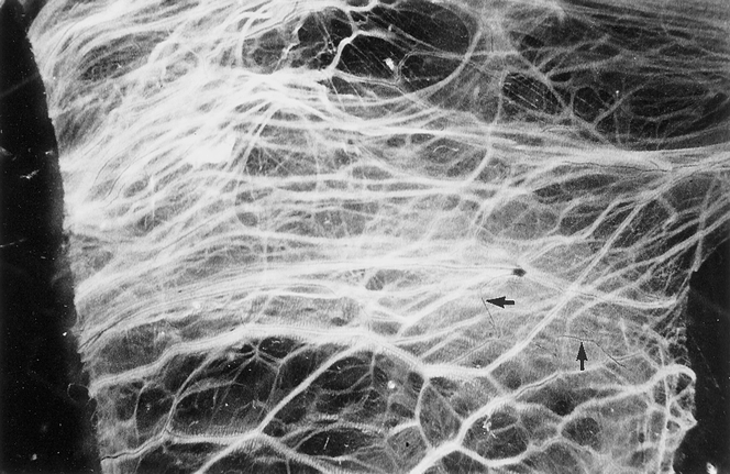
Figure 5. Distribution of blood vessels and nerve fibers in human peritoneal adhesions demonstrated in whole-mount preparations by acetylcholinesterase histochemistry. Generally, nerve fibers (black) accompanied blood vessels (white) arranged parallel to the long axis of the adhesion, but some nerve fibers branched independently (arrows) (×18).
