
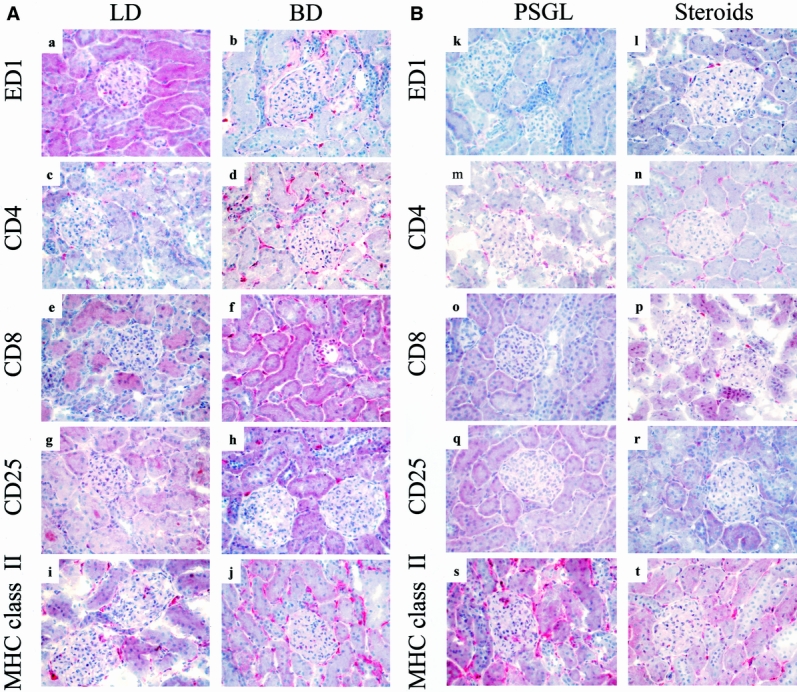
Figure 2. Effects of brain death on intragraft events 24 hours after transplantation. Numbers of ED1+, CD4+, CD8+, and CD25+ cells present in control kidneys are minimal (a–h). Donor brain death, in contrast, markedly enhanced the infiltration and accumulation of these leukocyte subtypes within perivascular and periglomerular areas (k–t). Cellular infiltrates were absent at 24 hours in the soluble P-selectin glycoprotein ligand (sPSGL) donor treatment group and in living donor kidneys, although their presence was diminished less by steroid treatment. Intragraft expression of MHC class 2 was increased and was particularly evident in interstitial macrophages in kidneys from untreated brain-dead donors; it was expressed only moderately in controls and after steroid treatment (i, j). sPSGL treatment did not affect MHC class 2 expression, which remained comparable to brain-dead organs (a–t). (Serial cryostat sections, representative of 4 animals per group per time-point; hematoxylin counterstain, ×400)