
Figure 6.
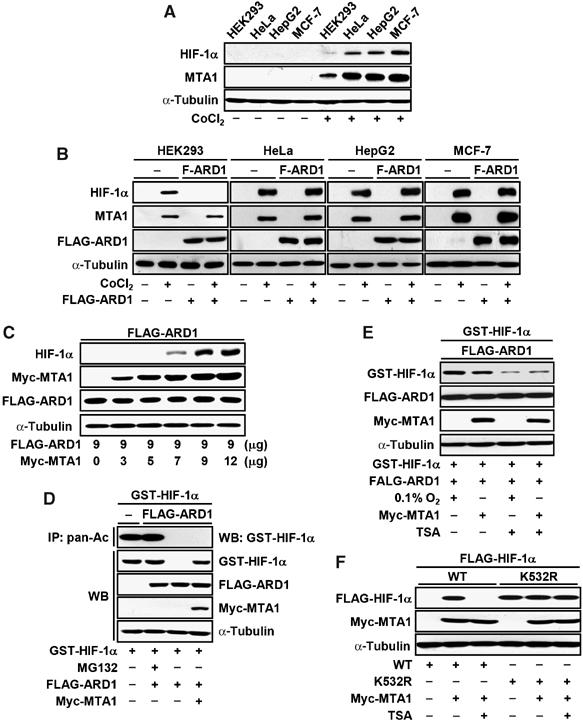
MTA1 counteracts to the ARD1-induced degradation of HIF-1α. (A) HEK293, HeLa, HepG2, and MCF-7 cells were treated with or without 100 μM CoCl2 for 24 h. The expression of HIF-1α, MTA1, and α-tubulin was analyzed by Western blot (WB) analysis. (B) HEK293, HeLa, HepG2, and MCF-7 cells were transfected with 3 μg of pCMV-Tag2C-FLAG-mARD1225 (F-ARD1) or empty vector. Transfected cells were incubated under hypoxia or normoxia for 24 h. The expression of HIF-1α, MTA1, FLAG-ARD1 (F-ARD1), and α-tubulin was analyzed by WB analysis. (C) HEK293 cells were transfected with 9 μg of pCMV-Tag2C-FLAG-mARD1225 (FLAG-ARD1) or the indicated amount of pCMV-Myc-MTA1. The expression of HIF-1α, Myc-MTA1, FLAG-ARD1, and α-tubulin was analyzed by WB analysis. (D) MCF-7 cells were transfected with 4 μg pEBG-HIF-1α (GST-HIF-1α), and 3 μg of each pCMV-Tag2C-FLAG-mARD1225 (FLAG-ARD1), pCMV-Myc-MTA1, or empty vector. Cells were treated with or without 10 μM MG132 for 1 h before being harvested. Whole-cell lysates (500 μg) were immunoprecipitated (IP) with anti-pan-Ac antibody, and then probed using anti-GST antibody. The expression of GST-HIF-1α, FLAG-ARD1, Myc-MTA1, and α-tubulin was analyzed by WB analysis. (E) MCF-7 cells were transfected with 4 μg pEBG-HIF-1α (GST-HIF-1α) with 3 μg of each pCMV-Tag2C-FLAG-mARD1225 or pCMV-Myc-MTA1 as indicated. After 1 h of transfection, cells were incubated under hypoxia or normoxia for 24 h as indicated. Cells were treated with or without 300 ng/ml TSA for 3 h before being harvested. The expression of GST-HIF-1α, FLAG-ARD1, Myc-MTA1, and α-tubulin was analyzed by WB analysis. (F) HEK293 cells were transfected with the indicated combinations of 1 μg of each p3XFLAGTM7.1-HIF-1α (WT), or p3XFLAGTM7.1-HIF-1α K532R (K532R), 3 μg of each Myc-MTA1 and empty vector. After 24 h of transfection, cells were incubated in the presence or absence of 300 ng/ml TSA for 3 h before being harvested. The expression of FLAG-HIF-1α, Myc-MTA1, and α-tubulin was analyzed by WB analysis.