
Figure 2.
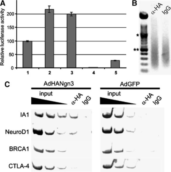
Ngn3 binds and activates the IA1 promoter. (A) The IA1 promoter is activated by Ngn3 and NeuroD1 in transient promoter–reporter assays in 293 cells. Cotransfection of an IA1 promoter–reporter construct with Ngn3 (2) or NeuroD1 (3) increases IA1 promoter activity by twofold as compared to the IA1 promoter only (1). The pGL3basic (4) and pGL3control (5) samples represent negative and positive controls for promoter activity, respectively. All experiments were performed in triplicate and repeated at least three times. (B, C) Ngn3 interacts with the 5′ flanking regions of the IA1 and NeuroD1 genes. (B) Ethidium bromide-stained agarose gel showing chromatin sonicated to an average length of 200–1000 bp. *1000 bp; **500 bp. (C) Chromatin immunoprecipitation with an anti-HA antibody or IgG was performed on chromatin derived from isolated human duct cells infected with either HANgn3 or GFP. DNA from input chromatin was serially diluted as a reference for semiquantitative PCR analysis. The figures are representative of four independent experiments. The IA1 gene promoter is coimmunoprecipitated in the HANgn3-transduced duct cells using an anti-HA antibody but not by addition of IgG. Similarly, NeuroD1, but not BRCA1 or CTLA-4, are specifically co-precipitated by the anti-HA antibody. IA1, NeuroD1, BRCA1 and CTLA-4 are not precipitated in the negative control cells transduced with AdGFP.