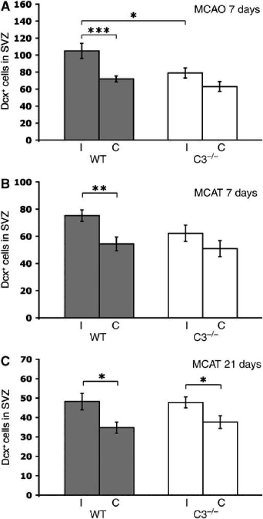
Figure 6.

Number of Dcxpos migrating neuroblasts in the SVZ in the ipsilateral (I) and contralateral (C) hemispheres of WT (n=7, 8, 8) and C3−/− (n=9, 7, 11) mice. The mice were subjected to focal cerebral ischemia by MCAO or MCAT. Dcxpos cells in the SVZ were counted in three sections per mouse 7 days after MCAO (A) and MCAT (B) and 21 days after MCAT (C). Values are the number of cells per section. *P<0.05, **P<0.01, ***P<0.005.
