
Figure 2.
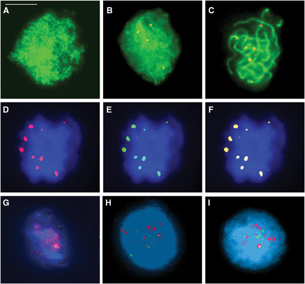
(A–C) Dual immunolocalization of AtMLH3 (red) and ASY1 (green). (A) At leptotene, ASY1 is localized to the developing chromosome axes. (B) AtMLH3 foci first become detectable during zygotene. (C) At pachytene 9–10, AtMLH3 foci are found in association with the chromosome axes. (D–F) Colocalization of the MutL homologues at pachytene. (D) AtMLH3 (red), (E) AtMLH1 (green) and (F) merged image. (G–I) Limited colocalization of AtMLH3 and AtMSH4. At early zygotene, AtMSH4 foci (red) are abundant with few AtMLH3 foci (green) detectable (G). At mid-zygotene (H) through to late zygotene/early pachytene (I), there is an increase in AtMLH3 foci and a reduction in AtMSH4 foci. Colocalization between the foci is consistently observed, but is limited to only few foci (1–2) per nucleus. Bar=10 μm.