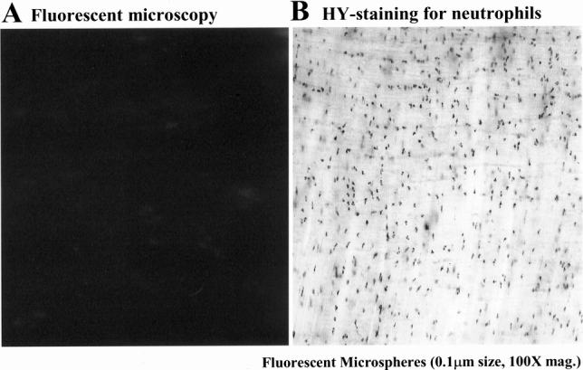
Figure 10. Subdiaphragmatic common lymph duct diversion prevented the appearance of fluorescent microspheres within phagocytes, which had extravasated into the manipulated jejunal muscularis. (Panel A) The muscularis whole mount from an animal in which the common subdiaphragmatic lymph duct was diverted by cutting after intestinal manipulation and microsphere injection showed the presence of no fluorescent beads in the muscularis (100× original magnification). However, these animals continued to have a strong leukocytic infiltrate, as shown by the presence of numerous myeloperoxidase-positive neutrophils within the manipulated muscularis (panel B, 200× original magnification).
