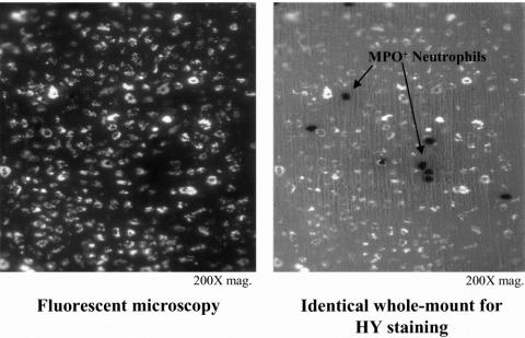
Figure 3. Double-labeling of this manipulated whole mount after 24 hours shows that the microsphere-laden cells in panel A photographed under fluorescent lighting are not identified as the strongly myeloperoxidase neutrophils shown in panel B, photographed with partial transilluminating light (200× original magnification).

An official website of the United States government
Here's how you know
Official websites use .gov
A
.gov website belongs to an official
government organization in the United States.
Secure .gov websites use HTTPS
A lock (
) or https:// means you've safely
connected to the .gov website. Share sensitive
information only on official, secure websites.
