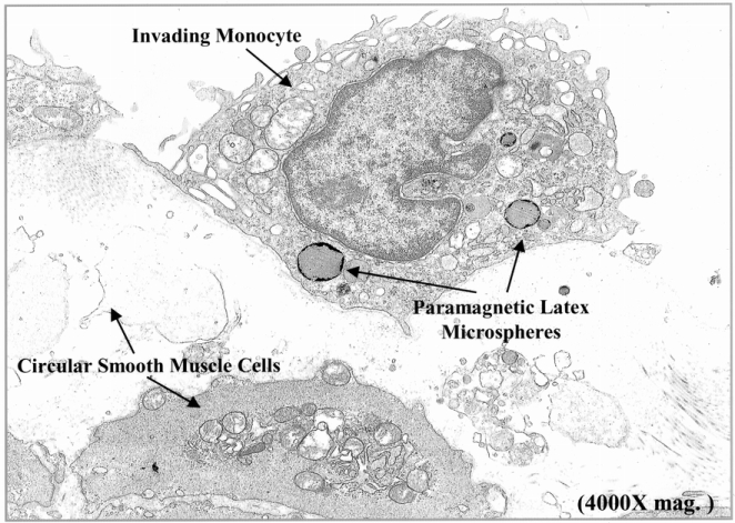
Figure 6. Electron microscopic confirmation of the microsphere-laden phagocytes as monocytes lying within the manipulated jejunal muscularis. Jejunal muscularis preparations fixed in glutaraldehyde for electron microscopy show the accumulation of 0.46-μm paramagnetic latex beads in phagocytes, which had structural cellular characteristics of monocytes. Each monocyte generally contained several microspheres and could be observed to be engulfing necrotic smooth muscle cells (4,000× original magnification).
