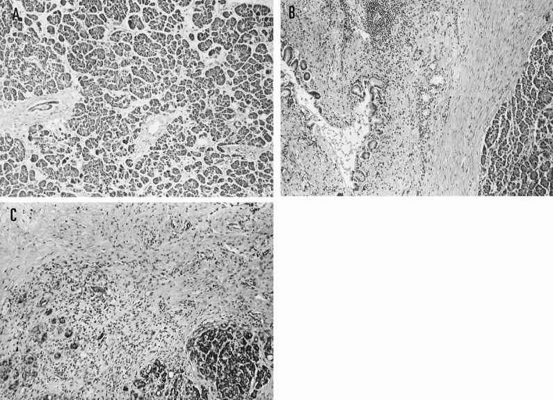
Figure 2. Surgical specimens of the pancreas in the three groups (hematoxylin and eosin, ×40). (A) No fibrosis group. Pancreatic edema and a few inflammatory cell infiltrations into the parenchyma of the pancreas are shown. (B) Periductal fibrosis group. Periductal fibrosis and many inflammatory cell infiltrations are seen. (C) Intralobular fibrosis group. Acinar necrosis, lobular fibrosis, and inflammatory cell infiltrations are seen.
