
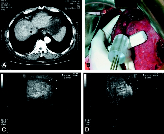
Figure 3. A 5-cm hepatocellular carcinoma at the dome of the liver (A, arrow) treated by intraoperative radiofrequency ablation using a clustered probe (B). Intraoperative ultrasound provides guidance to positioning of the probe (C, arrow shows the tip of the probe) in the tumor before starting radiofrequency ablation, but the exact margin of ablation is obscured by hyperechoic shadow resulting from thermal changes in the tissue after starting the ablation (D, arrows).