
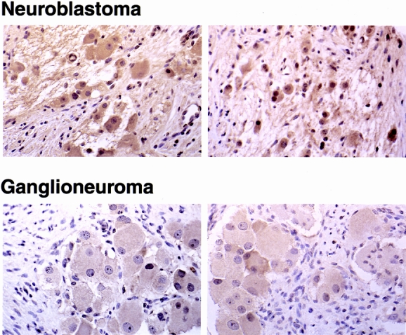
Figure 2. Expression of gastrin-releasing peptide (GRP) in neuroblastomas. Representative histologic sections of immunohistochemical staining with GRP antibody in neuroblastomas compared with ganglioneuromas (×100). Large mature ganglion cells show GRP protein (brown staining) in the cytoplasm.