
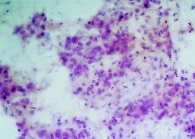
Figure 4. Suspicious for malignancy. This sheet of cells shows crowding, disorganization, nuclear pleomorphisms, and variation in the presence and size of nucleoli. Nuclear membranes are irregular as well. Hematoxylin and eosin, ×250 original magnification.