
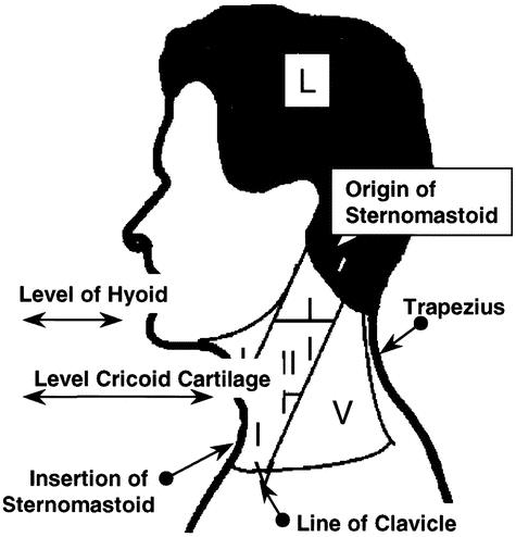
Figure 1. Schema for the location of abnormality when interpreting FDG-PET, CT, and histopathology. Levels are represented bilaterally and abbreviated as IR: level I on the right, IL: level I on the left, etc. They are defined as follows: Level I: (the anterior triangle) bounded by the anterior boarder of sternocleidomastoid, the inferior boarder of the mandible, and the midline anteriorly; level V (the posterior triangle) bounded by the posterior border of sternocleidomastoid, the superior boarder of the clavicle, the superior boarder of trapezius, and the midline posteriorly; and levels II, III, and IV, in relation to the upper, middle, and lower thirds of sternocleidomastoid. Landmarks for separating levels II, III, and IV are the level of the hyoid bone, between the upper and middle third and the level of the cricoid cartilage between the middle and lower thirds of sternocleidomastoid.