
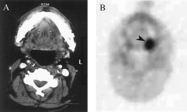
Figure 2. (A) CT scan of patient 25, which is negative for a primary left posterior glossal tumor. (B) FDG-PET transaxial scan showing the glossal primary tumor (black arrow). Pathology from surgery showed a 1.4 × 1.2 × 1.0-cm tumor at this site.
Official websites use .gov
A
.gov website belongs to an official
government organization in the United States.
Secure .gov websites use HTTPS
A lock (
) or https:// means you've safely
connected to the .gov website. Share sensitive
information only on official, secure websites.
Figure 2. (A) CT scan of patient 25, which is negative for a primary left posterior glossal tumor. (B) FDG-PET transaxial scan showing the glossal primary tumor (black arrow). Pathology from surgery showed a 1.4 × 1.2 × 1.0-cm tumor at this site.