
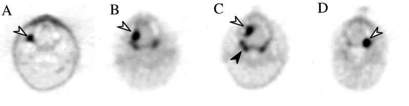
Figure 3. FDG-PET scans of primary tumors that were negative on CT scan. (A) Patient 1, with a right retromolar trigone oral cavity tumor (arrow). (B) Patient 33, with right-sided tongue tumor. (C) Patient 39, with a right anterior tongue tumor. (D) Patient 25, left oral cavity tumor. Normal FDG uptake in Waldeyer’s ring is evident (black arrow in C).