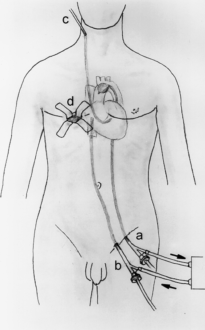
Figure 3. Typical operative setup for minimally invasive mitral valve repair at New York University Medical Center. (A) Femoral arterial cannulation with balloon catheter introduced for occlusion of the ascending aorta. (B) Femoral venous cannulation with tip of catheter positioned into the right atrium. (C) Percutaneous right internal jugular retrograde cardioplegia catheter positioned in the coronary sinus. (D) Right minithoracotomy incision performed via the inframammary crease through the fourth intercostal space.
