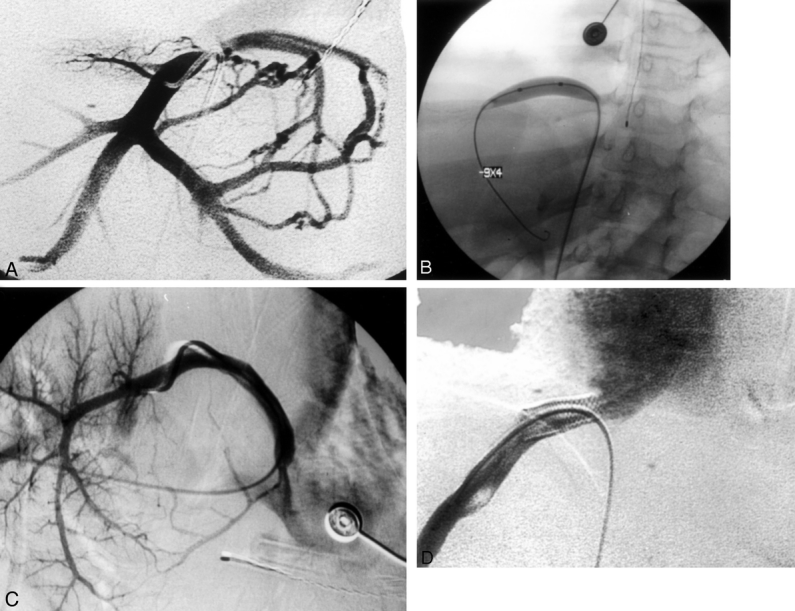
Figure 4. Hepatic vein stenosis demonstrated by venogram obtained with contrast injection into the inferior vena cava after a percutaneous femoral puncture. (A) Fluoroscopic image demonstrating venoplasty of stenotic segment. (B) Site of recurrent stenosis requiring stenting. (C) Placement of endovascular stent at stenotic point (D).
