
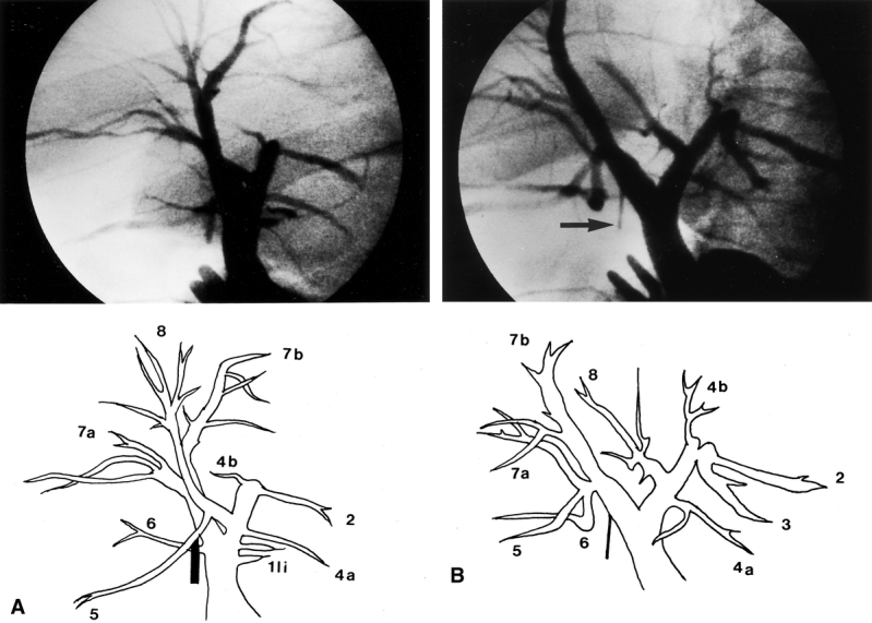
Figure 1. Operative cholangiogram of a 56-year-old donor. (A) This film was taken in an anteroposterior position. Interpretation was difficult because the liver was relatively small and had rotated into the right subphrenic cavity. (B) By rotating the x-ray tube to obtain a right antero-oblique view, two separate right hepatic ducts were clearly seen. The Liga clip (arrow) marks the proposed position of the division of the right anterior hepatic duct.