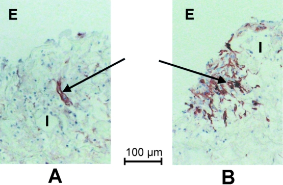
Figure 3. Procollagen type I positive fibroblasts in ePTFE tubes treated with 10 μg/mL GM-CSF (A) and control (B) from the same patient. E, external surface of ePTFE tube, I, intertrabecular space. Arrow indicates immunopositive fibroblast. Scale bar = 100 μm.
