
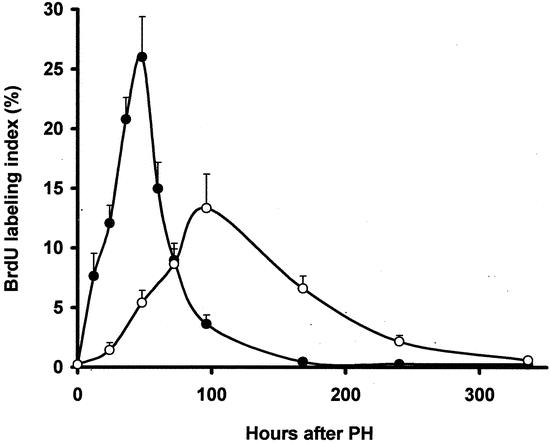
Figure 5. Effects of angiostatin on the proliferation of hepatocytes in resting and regenerating liver. BrdU uptake is represented by the ratio of labeled and nonlabeled hepatocytes times 100%. In normal resting liver the hepatocellular BrdU labeling index was approximately 0.2% (data not shown). Two days following 70% partial hepatectomy (PH group) a maximum of 26 ± 3.4% hepatocytes was labeled. In angiostatin-treated mice (PH/AS group) the BrdU labeling index peak was delayed to 96 hours after 70% partial hepatectomy. The BrdU peak was also significantly lower compared to the PH group (13.3 ± 2.9%;P < .001 vs. PH group).