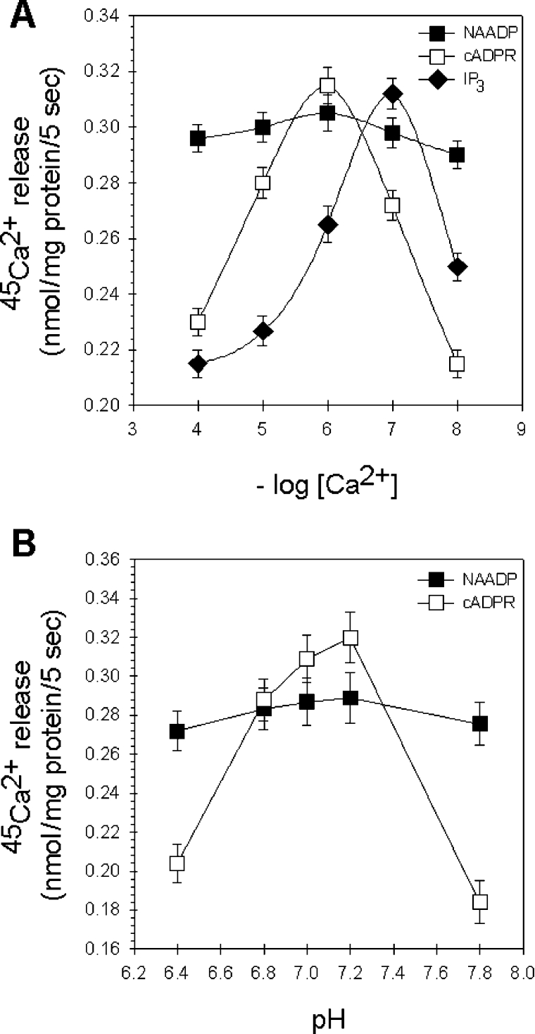
Figure 5. Ca2+- and pH-dependence of the NAADP-induced Ca2+ release.
(A) Extravesicular free Ca2+ concentration-dependence of the IP3-, cADPR- and NAADP-mediated system in passively loaded liver microsomes. Extravesicular pCa (4–8) was set by EGTA (200–750 μM), NAADP (■), IP3 (◆) and cADPR (□) were applied at supramaximal concentrations (10 μM). (B) Differential effect of pH on the cADPR- and NAADP-sensitive Ca2+-releasing system. The pH of the Ca2+-release medium was changed from 6.4 to 7.8, and the amount of 45Ca2+ released by 10 μM cADPR (□) and 10 μM NAADP (■) was determined.

