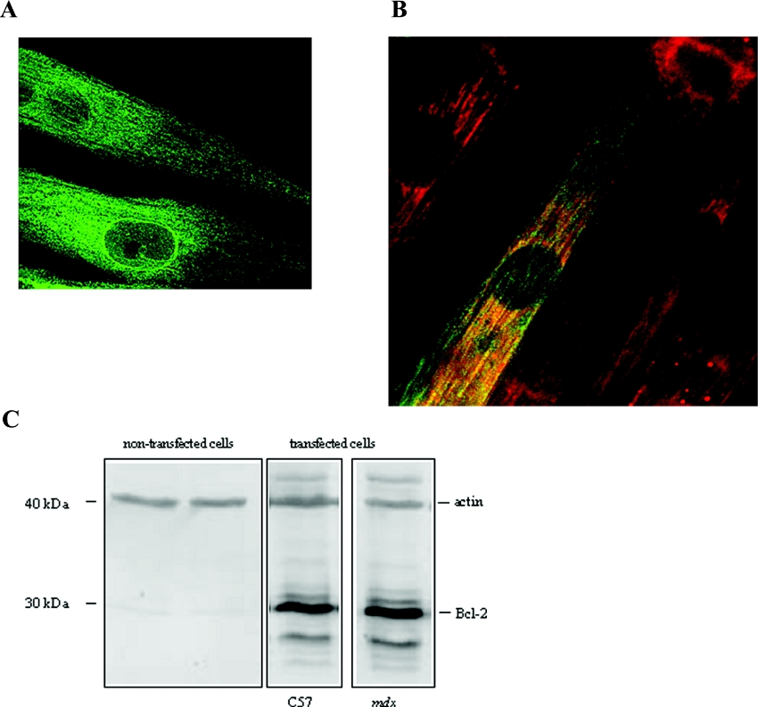
Figure 1. Bcl-2 localization and overexpression.
(A) Confocal section of mdx myotubes labelled with anti-Bcl-2 antibody for protein localization (green fluorescence). (B) Mitotracker (red fluorescence) and co-localization of mitochondria and Bcl-2 (yellow fluorescence). (C) Western-blot analysis of cell lysates from non-transfected and transfected control C57 and mdx myotubes. The blot was probed with anti-Bcl-2 antibody. Actin was used as control.

